
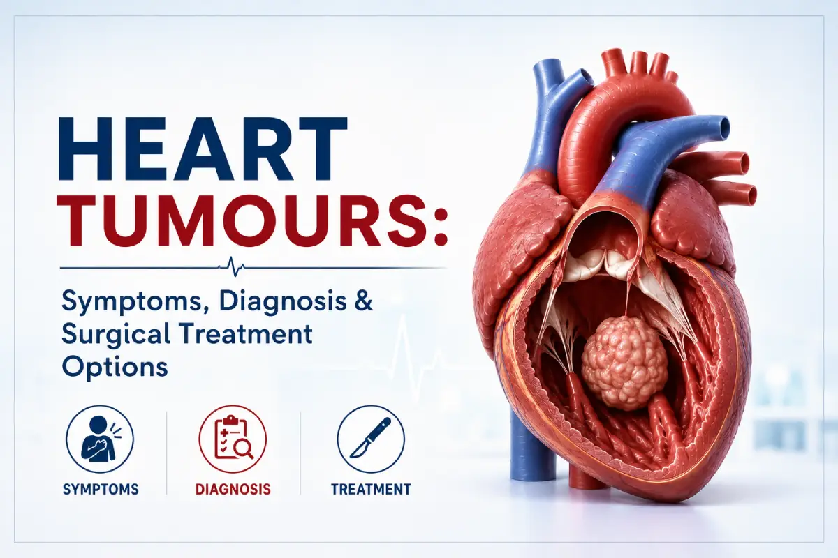
Heart Tumours: Symptoms, Diagnosis & Surgical Treatment Options
A heart tumour, also known as a cardiac tumour, is an abnormal growth that develops within the heart or nearby cardiac structures. Heart tumours are uncommon, and they can vary considerably in their type, location, size, behaviour, and effect on normal heart function.
Some cardiac tumours are benign, meaning they are not cancerous, while others are malignant. Tumours can originate within the heart itself or spread to the heart from cancer elsewhere in the body.
Even a benign heart tumour can sometimes become clinically important if its location interferes with blood flow, heart valves, normal heart rhythm, or other cardiac structures. For this reason, treatment decisions depend on much more than whether a tumour is classified as benign or malignant.
This guide explains the different types of heart tumours, possible symptoms, how cardiac tumours are diagnosed, and when surgical removal may be considered.

What Is a Heart Tumour?
A heart tumour is an abnormal mass involving cardiac tissue, the heart chambers, heart valves, or structures surrounding the heart.
Cardiac tumours can broadly be divided into:
- Primary cardiac tumours: tumours that begin within the heart.
- Secondary or metastatic cardiac tumours: tumours that have spread to the heart from another part of the body.
Primary cardiac tumours may be benign or malignant. Their clinical importance depends on their location, size, movement, growth pattern, and effect on circulation and heart function.
Are All Heart Tumours Cancerous?
No. A heart tumour does not automatically mean cancer.
Many primary cardiac tumours are benign. However, the term “benign” does not necessarily mean that the tumour can be ignored.
A non-cancerous tumour located inside a heart chamber may still interfere with blood flow, affect a heart valve, contribute to abnormal heart rhythms, or create other cardiovascular complications.
Malignant cardiac tumours behave differently and usually require a broader multidisciplinary treatment strategy.
What Is a Cardiac Myxoma?
A cardiac myxoma is a benign primary heart tumour and is one of the best-known types of primary cardiac tumour.
Myxomas commonly develop inside one of the upper chambers of the heart. Their exact effect depends on their size, attachment point, mobility, and location.
A myxoma may sometimes interfere with blood passing through a heart valve or chamber. In other cases, fragments associated with the tumour or surrounding material may travel through the circulation and create complications elsewhere in the body.
What Other Types of Heart Tumours Can Occur?
Several different cardiac tumour types are possible.
Examples of benign primary cardiac tumours can include:
- Myxoma
- Papillary fibroelastoma
- Lipoma
- Fibroma
- Rhabdomyoma
- Hemangioma
- Other uncommon benign cardiac masses
Primary malignant cardiac tumours are much less common and may include different forms of sarcoma and other rare cancers.
Cancers arising elsewhere in the body can also spread to cardiac or surrounding tissues. These are considered metastatic rather than primary heart tumours.
Where Can Heart Tumours Develop?
A cardiac tumour can occur in different parts of the heart.
Possible locations include:
- Left atrium
- Right atrium
- Left ventricle
- Right ventricle
- Heart valves
- Heart muscle
- Structures surrounding the heart
Location is extremely important because even a relatively small mass can cause significant problems if it obstructs an important blood-flow pathway or interferes with valve function.
What Symptoms Can a Heart Tumour Cause?
Symptoms vary significantly. Some cardiac tumours may be discovered unexpectedly during testing for another problem, while others cause noticeable cardiovascular symptoms.
Possible symptoms can include:
- Shortness of breath
- Reduced exercise tolerance
- Unexplained fatigue
- Palpitations
- Dizziness or light-headedness
- Fainting
- Chest discomfort
- Swelling of the legs or ankles
- Symptoms related to obstruction of blood flow
- Other symptoms depending on tumour location and behaviour
These symptoms are not specific to heart tumours and are much more commonly caused by other conditions. Proper medical evaluation is therefore essential rather than assuming that a symptom represents a cardiac mass.
Why Can the Location of a Heart Tumour Be Important?
The heart depends on unobstructed blood flow through its chambers and valves. A tumour located near one of these pathways can mechanically interfere with circulation.
For example, a mobile tumour within a chamber may move closer to a valve during parts of the cardiac cycle and potentially interfere with normal blood flow.
A tumour involving or located near cardiac electrical pathways may also contribute to abnormal heart rhythms in some patients.
Can a Heart Tumour Affect a Heart Valve?
Yes. Depending on its location, a cardiac mass can interfere with valve opening or closing.
This can produce functional obstruction or abnormal blood flow even if the valve tissue itself was originally normal.
Detailed echocardiography can help doctors understand how a mass interacts with nearby valves and cardiac structures.
Can Heart Tumours Cause Abnormal Heart Rhythms?
Some cardiac tumours can be associated with arrhythmias, particularly when they involve or affect cardiac tissue associated with the heart’s electrical system.
Patients may notice palpitations, irregular heartbeat, dizziness, weakness, or other symptoms.
However, abnormal rhythms have many more common causes, so appropriate cardiac evaluation is required.
Can a Cardiac Tumour Affect Blood Circulation?
Yes. Tumours can sometimes obstruct normal inflow or outflow through the heart or interfere with valve function.
Some cardiac tumours can also be associated with embolic complications if material travels through the bloodstream.
The significance of this risk varies according to the type, mobility, location, and other characteristics of the mass.
How Are Heart Tumours Diagnosed?
Cardiac tumours are generally evaluated with a combination of clinical assessment and cardiac imaging.
The diagnostic process may involve:
- Medical history and physical examination
- Electrocardiogram (ECG)
- Echocardiography
- Transesophageal echocardiography in selected cases
- Cardiac MRI
- CT imaging
- Blood investigations
- Other tests depending on the suspected tumour type
Why Is Echocardiography Important?
Echocardiography uses ultrasound to create images of the heart and can be particularly valuable when investigating a cardiac mass.
It can help doctors assess:
- Location of the mass
- Approximate size
- Mobility
- Attachment to cardiac structures
- Relationship with heart valves
- Effect on blood flow
- Heart chamber size
- Overall cardiac function
What Is Transesophageal Echocardiography?
Transesophageal echocardiography, often abbreviated as TEE or TOE, uses a specialized ultrasound probe positioned in the oesophagus to obtain detailed images of certain heart structures.
Because the oesophagus lies close to the heart, this approach can provide detailed views of some cardiac masses and their attachment points.
It is used when clinically appropriate rather than being required for every patient.
Why Is Cardiac MRI Used?
Cardiac MRI can provide detailed information about the location, size, structure, and tissue characteristics of a cardiac mass.
It can also help doctors understand how the tumour relates to surrounding heart structures and can be useful when planning treatment.
When Is a CT Scan Helpful?
CT imaging can provide detailed anatomical information about the heart, major blood vessels, surrounding chest structures, and selected tumour characteristics.
It may be used alone or together with other imaging methods depending on the clinical situation.
Can Imaging Tell Whether a Heart Tumour Is Benign or Malignant?
Imaging can provide important clues about the nature of a cardiac mass, including its location, growth pattern, tissue characteristics, and whether surrounding structures appear involved.
However, imaging does not always provide absolute certainty about tumour type.
The final diagnosis may depend on the complete clinical picture, surgical findings, and laboratory analysis of removed tissue when surgery is performed.
Does Every Heart Tumour Need Surgery?
No. Treatment depends on the tumour type, location, size, symptoms, potential complications, and whether the mass is believed to be benign, malignant, or metastatic.
Possible management strategies may include:
- Careful monitoring in selected situations
- Surgical removal
- Treatment directed toward an underlying cancer
- Oncology treatment for malignant disease
- Management of associated cardiac complications
- A multidisciplinary treatment strategy
When May Heart Tumour Surgery Be Recommended?
Surgical removal may be considered when a cardiac tumour is causing or has the potential to cause clinically important problems.
Factors influencing the decision can include:
- Tumour type
- Location within the heart
- Size
- Mobility
- Symptoms
- Obstruction of blood flow
- Interference with heart valves
- Risk of embolic complications
- Effect on heart rhythm or function
- Likelihood that the tumour can be removed safely
- Overall health of the patient
Why Are Some Benign Heart Tumours Removed?
A benign tumour does not spread to distant organs in the way a malignant cancer can, but its location inside the heart can still make it dangerous.
For example, a benign tumour may interfere with normal blood flow or valve function or create a risk of other cardiac complications.
For this reason, surgical removal may be recommended for selected benign cardiac tumours even when they are not cancerous.
How Is Heart Tumour Surgery Performed?
The exact operation depends on the tumour’s location, size, attachment, and relationship with nearby heart structures.
The primary surgical objective is generally to remove the mass safely while preserving normal cardiac structures and function whenever possible.
The surgeon may also need to repair structures affected by the tumour depending on the individual case.
Can Heart Tumour Surgery Be Minimally Invasive?
Selected patients with suitable tumour anatomy may potentially be considered for minimally invasive cardiac surgery using smaller surgical access points.
Suitability depends on several factors, including:
- Location of the tumour
- Size of the mass
- Attachment point
- Relationship with valves and other structures
- Need for additional cardiac procedures
- Previous cardiac surgery
- Overall patient health
Conventional cardiac surgery may still provide the safest and most effective access for complex tumours.
Can Robotic Surgery Be Used for Cardiac Tumours?
Robotic cardiac surgery may be an option for selected cardiac procedures when the anatomy and tumour characteristics allow safe access.
Robotic technology allows the surgeon to control specialized instruments through smaller access points.
However, cardiac tumour surgery must be planned according to the tumour rather than according to a preference for a particular technology. A robotic or minimally invasive technique is only appropriate when it allows safe and effective treatment.
Heart Tumour Treatment: Key Factors
| Factor | Why It Matters |
|---|---|
| Tumour Type | Helps determine whether monitoring, surgery, oncology treatment, or another strategy is appropriate |
| Location | Determines which heart structures may be affected and influences surgical access |
| Size | May influence obstruction, symptoms, and complexity of treatment |
| Mobility | May influence interaction with valves and other potential complications |
| Heart Function | Helps assess the effect of the tumour and overall treatment risk |
| Overall Health | Important when evaluating surgery and recovery |
| Final Treatment Plan | Individualized after detailed imaging and specialist assessment |
What Happens After Heart Tumour Surgery?
After surgery, the patient is monitored closely while heart rhythm, circulation, blood pressure, breathing, pain control, and overall postoperative recovery are assessed.
The recovery process depends on the operation performed and whether conventional, minimally invasive, or another surgical approach was used.
As recovery progresses, care generally focuses on:
- Heart and circulation monitoring
- Pain control
- Breathing exercises when recommended
- Wound care
- Gradual mobility
- Medication management
- Preparation for discharge
How Long Does Recovery Take After Heart Tumour Surgery?
There is no single recovery timeline for cardiac tumour surgery.
Recovery can depend on:
- Type of surgical approach
- Location and size of the tumour
- Complexity of removal
- Whether additional cardiac repair was required
- Age
- Heart function
- Overall medical health
- Postoperative complications
- Individual healing response
The cardiac surgical team provides individualized guidance regarding walking, lifting, driving, work, exercise, wound care, and follow-up.
What Happens to the Tumour After Removal?
When a cardiac tumour is surgically removed, the tissue may be examined by pathology specialists.
This analysis helps determine the exact type of tumour and provides information that may influence follow-up or additional treatment.
Can a Heart Tumour Come Back After Surgery?
Recurrence depends on the specific tumour type and individual circumstances.
Some cardiac tumours can recur after treatment, which is one reason follow-up imaging may be recommended even after apparently successful removal.
The frequency and duration of monitoring should be individualized according to the final diagnosis.
What Follow-Up May Be Needed?
Follow-up after treatment may include:
- Clinical examination
- Echocardiography
- Cardiac MRI or CT when appropriate
- Heart rhythm assessment
- Review of symptoms
- Monitoring heart function
- Pathology review
- Additional specialist care if required
What If the Cardiac Tumour Is Malignant?
Malignant cardiac tumours require individualized specialist management.
Treatment planning may involve cardiac surgeons, cardiologists, oncologists, imaging specialists, pathologists, and other healthcare professionals.
The treatment strategy depends on the tumour type, extent of disease, involvement of surrounding structures, overall health, and whether surgery is likely to provide benefit.
What Are Metastatic Heart Tumours?
A metastatic cardiac tumour develops when cancer that began elsewhere in the body spreads to the heart or surrounding cardiac structures.
This is different from a primary malignant cardiac tumour that originates within the heart itself.
Management usually focuses on the underlying cancer and the way cardiac involvement is affecting the patient.
Questions to Ask Your Cardiac Surgeon
If a cardiac mass or tumour has been identified, useful questions may include:
- Where exactly is the tumour located?
- How large is it?
- Does it appear benign or malignant?
- Could it be a cardiac myxoma?
- Is it affecting a heart valve?
- Is blood flow through the heart being obstructed?
- Do I need additional imaging?
- Is cardiac MRI required?
- Does the tumour need to be removed?
- What happens if it is monitored instead?
- Which surgical approach would be appropriate?
- Am I suitable for minimally invasive surgery?
- Could robotic surgery be considered?
- Will the removed tissue be examined?
- What follow-up will be necessary after treatment?
Frequently Asked Questions About Heart Tumours
Are heart tumours common?
Primary heart tumours are uncommon. Cardiac masses can represent several different conditions, so specialist imaging and evaluation are needed to establish the diagnosis.
Does a heart tumour mean cancer?
No. Primary heart tumours may be benign or malignant. A cardiac mass therefore should not automatically be assumed to represent cancer.
What is the most common benign primary heart tumour?
Cardiac myxoma is one of the most commonly recognized benign primary cardiac tumours.
Can a benign heart tumour still be dangerous?
Yes. A benign cardiac tumour can still cause important problems because of its location, particularly if it interferes with blood flow, heart valves, rhythm, or other cardiac structures.
Can an echocardiogram detect a heart tumour?
Echocardiography can identify many cardiac masses and provide important information about their location, size, mobility, and effect on surrounding structures.
Why might cardiac MRI be needed?
Cardiac MRI can provide additional detail about a mass and surrounding tissue and can help characterize the tumour and plan treatment.
Does every cardiac myxoma require surgery?
Treatment decisions must be individualized. Surgical removal is commonly considered for cardiac myxomas because their location and mobility can create clinically important complications.
Can heart tumour surgery be minimally invasive?
Selected cardiac tumours may potentially be treated through minimally invasive approaches when the anatomy and surgical requirements make this safe and appropriate.
Can a heart tumour return after removal?
Some tumour types can recur, although the risk varies. Follow-up imaging may therefore be recommended according to the final diagnosis.
Why Specialist Evaluation Matters
Finding a mass inside the heart can naturally create concern, but the term “heart tumour” includes several different conditions with very different behaviours and treatments.
Detailed cardiac imaging can help determine where the mass is located, how it interacts with surrounding structures, and which treatment options should be considered.
For selected tumours, surgical removal may provide definitive treatment. Other cases may require monitoring, oncology treatment, or multidisciplinary management.
The most appropriate treatment is determined by the exact tumour type, its location and behaviour, its effect on the heart, and the individual patient’s overall clinical condition.
Consult Dr. Arunansu Dhole for Heart Tumour Surgery
Dr. Arunansu Dhole, MBBS, DNB, MCh (CTVS), MRCS (Eng), is a Cardiothoracic & Vascular Surgeon providing advanced cardiac surgical care, including heart tumour surgery, minimally invasive cardiac surgery, robotic cardiac surgery, heart valve surgery, and coronary artery bypass surgery.
If a cardiac mass or heart tumour has been identified on echocardiography, CT, cardiac MRI, or another investigation, a detailed cardiothoracic surgical evaluation can help determine whether monitoring, surgical removal, minimally invasive treatment, or another approach may be appropriate.
Schedule a consultation with Dr. Arunansu Dhole for individualized evaluation and discussion of cardiac tumour diagnosis and surgical treatment options.
Important Medical Information
This article is intended for general patient education only and should not be used to diagnose a cardiac tumour or determine whether surgery is required. A cardiac mass may represent different conditions, and treatment depends on imaging findings, tumour characteristics, symptoms, pathology, heart function, and overall health. Please consult qualified healthcare professionals for individualized diagnosis and treatment recommendations.